Please activate JavaScript!
Please install Adobe Flash Player, click here for download
ePaper created 2017-05-12, 09:42:21 | version 1.38.0
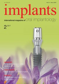
case report | Fig. 2: Description of the surgical procedure: a) clinical condition of the alveolar ridge that shows the edentulous area of teeth 14, 15 and 16 prior the beginning of the surgical procedure; b) crestal incision and blood collection with a sterile syringe for further bone graft; c) pilot drilling at the site of dental implantation with simultaneous ISL; d) 2.5 mm latch reamer with no cutting edge to start osteotomy widening at the crestal bone; e) 2.5 mm latch ream- er perforation in the bone starting the osseous widening; f) X-ray show- ing RBH (4.24 mm) and also 2.5 mm latch reamer insertion into the bone (4.29 mm). implants 2 2017 21 Fig. 2a Fig. 2b Fig. 2c Fig. 2d Fig. 2e Fig. 2f used (5 mm) in comparison with non-grafted sites (3 mm, P < 0.05), this may not be required to promote endo-sinus gain. Although there’s no consensus whether bone graft should be placed via ISL procedure or not, this option is highly recommended due to the benefits regarding osseous level maintenance. The aim of this study was to describe radiographic parameters of minimally-invasive internal sinus elevation in com- bination with plateau-form short implants. Thus this paper is intended to describe the surgical technique of a predictable procedure we developed during our 20 years of practical hands-on training with stu- dents and course participants. Case report A female 58-year-old patient consulted us because of her desire of functional and aesthetic repairment. The patient did not relate any medical background of clinical interest. She also signed informed consent prior the beginning of study, held ASA I status and re- quired dental implantation in the upper posterior maxilla with at least 4 mm on RBH and measured through a digital periapical radiograph by a single op- erator calibrated for this (Intraclass Correlation Coef- ficient: 0.83). Amoxicillin 500 mg was prescribed to the patient two days prior the surgical procedure, once every eight hours in order to avoid infections. Surgical pro- cedure was performed by a different trained clinician with more than 25 years of experience in the field. Surgical Technique Infiltrative anaesthesia was used in the procedure. Initially, a non-epinephrine anaesthesia was used (PRICANEST 4 %, Ropsohn Therapeutics, Bogotá, Colombia) in order to collect blood (Fig. 2b) to mix with the grafting material (50–500 µm Synthograft, Bicon Dental Implants, Boston, USA). Then 2 % Xylo- caine (Dentsply Pharmaceutical, York, USA) was used to complete the surgical procedure. Intrasulcular incision was performed by using a size 15 blade in a Bard-Parker scalpel. Full thickness flap was obtained in the area and then a previously pub- lished protocol8 was followed to perform ISL: pilot (2 mm diameter) drilling to achieve cortical perfora- tion was started. Pilot drilling length (1–2 mm) was determined upon residual bone height prior measure-