Please activate JavaScript!
Please install Adobe Flash Player, click here for download
ePaper created 2017-03-17, 12:41:55 | version 1.38.0
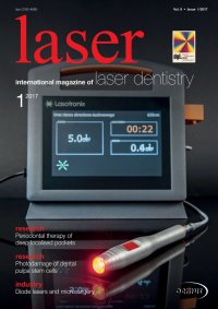
| research Photodamage of dental pulpa stem cells during 700 fs laser exposure Authors: Prof. Dr Karsten König & Dr Anton Kasenbacher, Germany Material and methods Cells Human stem cells from the dental pulp of adults (given by S. Gronthos, NIH, Bethesda, USA) were cultivated in a-modifi ed Eagle’s Medium (MEM) by Gibco BRL Life Technologies (Paisley, Scotland) while adding 20 % FCS, 2 mM L-glutamine, 100 µM L-ascorbate-2-phosphate, 100 u Penicillin and 100 µg/ml Streptomycin at 5 % CO2 and 37°C in 25 ml and 75 ml cell culture fl asks (Greiner, Frickenhausen, Germany).1 The cells were treated for experiments and cultivation with 0.25 % trypsin, 5 mM glucose, 0.05% EDTA in PBS for 5 minutes at 37 °C. After hav- ing been thus detached from the base of the culture fl asks, the cells were incubated in cell chambers (MiniCeM, JenLab GmbH, Jena, Germany) for laser (MiniCeM, JenLab GmbH, Jena, Germany) for laser microscopy. microscopy. Fig. 1: Femtosecond laser system with pulse-stretching unit. Laser-scanning microscope Sensor diode Objective Comparative studies were conducted on Chinese Comparative studies were conducted on Chinese hamster ovary (CHO) cells, which are available in hamster ovary (CHO) cells, which are available in many international laboratories as reference cells. many international laboratories as reference cells. Mirrors with three- point support Beamer expander Auto- correlator Grating 2 on motorized translation stage Pulse strechter Grating 1 Mirror 18 laser 1 2017 The CHO cells were incubated in Dulbeccos HAM-F12 Medium (Gibco BRL) with 10 % FCS, L-glutamine and an antibiotic mix of Penicillin, Streptomycin and Am- photericin B at 5 % CO2 and 37 °C. Trypsinisation cor- responded to that of the stem cells. Individual cells were marked by special diamond engravings in the exterior glass window. In case of the detection of cellular damage, these engravings were easily located by applying the phase-contrast technique. Laser microscopy An 80 MHz Ti:Sa Laser, Mai Tai (Spectra-Physics, Mountain View, USA) was applied for femtosec- ond-laser microscopy in the near infrared (NIR) spectrum. The laser power at microscope entrance spectrum. The laser power at microscope entrance and objective plane (power at the sample) were de- and objective plane (power at the sample) were de- termined by the measuring instrument Fieldmaster termined by the measuring instrument Fieldmaster (Coherent, Santa Clara, USA) and the measuring (Coherent, Santa Clara, USA) and the measuring head LM2 and varied by grey fi lters when necessary. head LM2 and varied by grey fi lters when necessary. The measured values were specifi ed as corrected in The measured values were specifi ed as corrected in the presented protocol. This correction results from the presented protocol. This correction results from a limited measurement area and altered radiation a limited measurement area and altered radiation conditions in the medium when compared to air. conditions in the medium when compared to air. Laser microscopy was realised via a modifi ed Laser microscopy was realised via a modifi ed LSM 410 (ZEISS, Jena, Germany) with a 40 x/1.3 LSM 410 (ZEISS, Jena, Germany) with a 40 x/1.3 oil immersion objective. The microscope scan oil immersion objective. The microscope scan modus 512 x 512 with a laser scan time t = 16 s was ap- plied for cell irradiation. The cells were scanned ten times at the same focus plane. These experiments were realised at a central wave- length of 800 nm. After ir radiation, the cells were Fs laser Attenuator