Please activate JavaScript!
Please install Adobe Flash Player, click here for download
ePaper created 2017-12-06, 12:03:50 | version 1.38.0
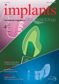
| case report Fig. 6 Fig. 7 Fig. 8 Case 1—Fig. 6: Maxilla with multi-unit abutments. Fig. 7: Basal view of the screw-retained bridge. Fig. 8: Coronal view of the screw-retained bridge. mill the required geometry or there was an insuffi- cient conical connection surface between the framework and implant to eliminate transversal forces leading to screw loosening (Table 4, see QR Code Tables). The patient needed to be able to brush and floss each implant easily, giving consideration to the fact that all of the patients in this series were over 60 years old, with an average age of 65 to 70, and some were already in their 80s. CAD/CAM-milled superstructure The digital era of dentistry has reached our practice. A digital workflow is not possible without compro- mises. Nevertheless, new materials, manufacturers and systems are easily integrated into any workflow or milling system. Also, anatomical findings can often avoid techniques like intraoral scanning. In this case series, only six cases were performed with intraoral scanning. Owing to the very high soft-tissue quantity that we saw in many cases, scan bodies were not always sufficient for the scanners to perform digital impressions. The matching of an edentulous jaw with a non-edentulous one is possible with additional aid. Even in cases that were scanned intraorally, we manufactured 3-D models to proceed with the jaw matching. Case 1 In the first case, a female patient with a partially edentulous maxilla was treated. All of the remaining teeth had to be extracted. One implant was an imme- diate implantation. The patient received a removable provisional prosthesis for two months. Owing to the implant surface of the manufacturer, we decided to re-enter after eight weeks. The final loading took place 12 weeks after implant placement. Although one implant had no optimal angulation, a screw-re- tained bridge was possible. The prosthesis received a resin layer facing the ceramic bridges in the mandible. The replacement of the missing teeth with a pros- thesis was sufficient for optimal support of the lower face soft tissue. Furthermore, a welcome side effect of such treatment is the decrease of wrinkles through the soft-tissue support. With this, the patient looked ten years younger. A very important focus of the prosthetic rehabilitation was hygiene maintenance. The prostheses were loaded on multi-unit abut- ments. Although we generally load maxillae at the im- plant level, the implant system used in this case was very new and milling centres were not able to mill the internal geometry or use other cast scan bases. Nevertheless, loading maxillae at the implant level re- duces weak points at the superstructure level. Here, we would have six screws that could loosen. Since the maxilla is not mobile, there are no torsional forces on the implants or the superstructure (Figs. 2–12). Case 2 In the second case, treatment of a mandible was performed. All of the teeth needed to be extracted; however, initially, three molars were retained for the stability of the provisional prosthesis. Owing to emo- tional reasons, the last two molars were extracted some months after loading the implants. Four of the five implants were placed immediately. The fifth one, placed in region #46, was a late implantation. We could clearly see that, at the point of loading, there was slight vertical bone resorption at #46. This was not true resorption, but more the establishment of a new biological width. This new biological width is inevitable. In late defects, biological width is lower if plat- form-switched implants are used and placed cor- rectly and higher if other platforms are used. Imme- diate implantations seem to behave more predictably: the transformation of the socket begins with the implant placement and not after the re-entry. With this, we see no vertical bone loss in the first weeks or months after loading the implants. The long-term stability is not affected by the new biological width. The main factors for long-term stability are bone quantity, soft-tissue quantity and quality, prosthetic and masticatory forces, as well as proper oral hygiene at home (Figs. 13–19). 14 implants 4 2017