Please activate JavaScript!
Please install Adobe Flash Player, click here for download
ePaper created 2018-04-06, 15:48:03 | version 1.38.0
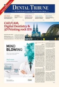
10 mCME Dental Tribune Middle East & Africa Edition | 3/2017 The treatment of traumatic dental injuries CAPP designates this activity for 1 CE Credits mCME articles in Dental Tribune have been approved by: HAAD as having educational content for 1 CME Credit Hours DHA awarded this program for 1 CPD Credit Points Fig. 1a: Clinical case of two uncomplicated crown frac- tures in which the two broken pieces were located and reattached. (Photos/Provided by American Association of Endodontists) Figs. 1b, c: After the two pieces had been attached, a chamfer was cut along the fracture line and additional compos- ite cured in place. This will both increase the strength of the attachment and better hide the fracture line. By Dr Asgeir Sigurdsson, USA When treating dental trauma, the timeliness of care is key to saving the tooth in many cases. It is, therefore, important for all dentists to have an understanding of how to diagnose and treat the most common dental injuries. This is especially critical in the emergency phase of treatment. Proper management of dental trau- ma is most often a team effort with general dentists, pediatric dentists or oral surgeons on the front line of the emergency service, and endodontic specialists joining the effort to pre- serve the tooth with respect to the pulp, pulpal space and root. An in- formed and coordinated effort from all team members ensures that the patient receives the most effi cient and effective care. Recently, a panel of expert members of the American Association of En- dodontists prepared an updated ver- sion of Guidelines for the Treatment of Traumatic Dental Injuries.1,2 These guidelines were based, in part, on the current recommendations of the International Association of Dental Traumatology (see www.iadtdental- trauma.org for more information). This article provides an overview of the AAE guidelines; the complete guidelines are available for free download at www.aae.org/clinical- resources/trauma-resources.aspx. The benefi t of adhering to guide- lines for treatment of dental trauma was recently shown in a study by Bucher et al.3 The study found that, compared with cases treated with- out compliance to guidelines, cases that adhered to guidelines produced more favorable outcomes, includ- ing signifi cantly lower complication rates. The study also found that early follow-up visits were essential to en- sure prompt treatment of complica- tions when they arose.3 Emergency care Prior to any treatment, one must evaluate the injury thoroughly by careful clinical and radiographic in- vestigation. 1 1/2 to 2 mm high-speed diamond bur with copious water cooling Fig. 2a: Schematic diagram of minimal pulpotomy, where an approximately 2-mm reservoir is cut with a high- speed diamond bur and copious water cooling and calcium hydroxide mixed with sterile water placed. (Schematic drawings/Provided by Dr. Sigurdsson) Pulp capping agent Glass-ionomer Fig. 2b: Glass ionomer or a protective liner is placed over the pulp capping agent to ensure it stays in place during etching and bonding. It is recommended to follow a check list to ensure that all necessary infor- mation regarding the patient and the injury is gathered, including: 1. Patient’s name, age, sex, address and contact numbers (include weight for young patients). 2. Central nervous system symp- toms exhibited after the injury. 3. Patient’s general health. 4. When, where and how the injury occurred. 5. Treatment the patient received elsewhere. 6. History of previous dental injuries. 7. Disturbances in the bite. 8. Tooth reactions to thermal chang- es or sensitivity to sweet/sour. 9. If the teeth are sore to touch or during eating. 10. If the patient is experiencing spontaneous pain in the teeth. Once all of this information is gath- ered, a diagnosis can be made and appropriate treatment rendered. If the injured individual is not a patient of record, all necessary demographic information should be gathered as soon as the patient arrives and prior to any assessment. In the case of avulsion and the tooth being out of its socket, one should immediately place the tooth in a physiological solution of specialized media (such as Hank’s Balanced Salt Solution) or milk, or saline if those are not available. Only after the tooth is secured in solution should one ob- tain the patient’s information. Once the patient is seated in the dental chair, it is necessary to do a quick central nervous system (CNS) evalu- ation before proceeding with further assessments. Often, the dentist is the fi rst health- care provider to see the patient after a head injury (any dental trauma is, by defi nition, a head injury) and must assess the risk of concussion or hemorrhage. It has been estimated by a meta-analysis that the preva- lence of intracranial hemorrhage after a mild head injury is 8 percent, and the onset of symptoms can be delayed for minutes to hours.4 The Table 1. Follow-Up Procedures for Fractured Permanent Teeth and Alveolar Fractures Fig. 2c: Clinical pictures of the minimal pulpotomy. most common signs of serious cer- ebral concussion or hemorrhage are loss of consciousness or post-trau- matic amnesia. Nausea/vomiting, fl uids from the ear/nose, situational confusion, blurred vision or uneven pupils, and diffi culty of speech and/ or slurred speech may also indicate serious injury.5 Once the patient has been cleared of any CNS issues, the dental trauma should be assessed. The key is to ob- tain comprehensive information about the injury and, to do so, one must conduct thorough extra-oral and intraoral clinical exams as well as appropriate radiographic evalua- tions. The new AAE guidelines recommend taking one occlusal and two periapi- cal radiographs with different lateral angulations for all dental injuries, including crown fractures. If cone- beam computed tomography is available, it should be considered for more serious injuries, such as crown/ root, root and alveolar fractures, as well as all luxation injuries. ÿPage 11 TIME 4 Weeks 6-8 Weeks 4 Months 6 Months 1 Year Yearly for 5 Years Crown Fracture Crown-Root Fracture Uncomplicated Complicated Uncomplicated Complicated Root Fracture Alveolar Fracture Splint removal*, clinical and radiographic control Splint removal and clinical and radiographic controls Clinical and radiographic control Clinical and radiographic control Clinical and radiographic control Clinical and radiographic control Clinical and Clinical and radiographic radiographic control control Clinical and radiographic control Clinical and radiographic control Clinical and radiographic control Clinical and radiographic control Splint removal**, clinical and radiographic control Clinical and radiographic control Clinical and Clinical and radiographic radiographic control control Clinical and Clinical and radiographic radiographic control control Clinical and Clinical and radiographic radiographic control control mCME SELF INSTRUCTION PROGRAM CAPPmea together with Dental Tribune provides the opportunity with its mCME - Self Instruction Program a quick and simple way to meet your continuing education needs. mCME offers you the fl exibility to work at your own pace through the material from any location at any time. The content is international, drawn from the upper echelon of dental medicine, but also presents a regional outlook in terms of perspective and subject matter. Membership Yearly membership subscription for mCME: 1,100 AED One Time article newspaper subscription: 250 AED per issue. After the payment, you will receive your membership number and allowing you to start the program. Completion of mCME • • • • • • • • mCME participants are required to read the continuing medical education (CME) articles published in each issue. Each article offers 2 CME Credit and are followed by a quiz Questionnaire online, which is available on www.cappmea.com/ mCME/questionnaires.html. Each quiz has to be returned to events@cappmea.com or faxed to: +97143686883 in three months from the publication date. A minimum passing score of 80% must be achieved in order to claim credit. No more than two answered questions can be submitted at the same time Validity of the article – 3 months Validity of the subscription – 1 year Collection of Credit hours: You will receive the summary report with Certifi cate, maximum one month after the expiry date of your membership. For single subscription certifi cates and summary reports will be sent one month after the publication of the article. The answers and critiques published herein have been checked carefully and represent authoritative opinions about the questions concerned. Articles are available on www.cappmea.com after the publication. For more information please contact events@cappmea.com or +971 4 3616174 FOR INTERACTION WITH THE AUTHORS FIND THE CONTACT DETAILS AT *Splint removal in apical third and mid-root fractures; **Splint removal with a root fracture near the cervical area THE END OF EACH ARTICLE. (Tables/Provided by American Association of Endodontists)